arthroscopy
Home
Our Expertise
Arthroscopy & Sports Injury Treatment
Arthroscopy is a minimally invasive surgical procedure used to diagnose and treat problems inside a joint. It is commonly known as "Pinhole Surgery" because it requires only tiny incisions, resulting in less pain, minimal tissue damage, and faster recovery.
What is Arthroscopy?
During arthroscopy, a specialized instrument called an arthroscope is inserted into the joint through a small incision. The arthroscope contains a camera and light source that allow the surgeon to view the inside of the joint on a high-definition monitor.
Most arthroscopic procedures are performed as day-care surgeries, allowing patients to return home the same day or after a short hospital stay.
Minimally Invasive
Tiny incisions reduce tissue damage and surgical trauma.
Faster Recovery
Most patients recover quicker compared to traditional surgery.
Accurate Diagnosis
Provides a clear view of joint structures for precise evaluation.
Less Postoperative Pain
Smaller incisions result in less discomfort after surgery.
Main Indications for Arthroscopy
- Meniscal Tears
- Ligament Injuries (ACL, PCL)
- Articular Cartilage Damage
- Loose Bodies Inside the Joint
- Intra-Articular Fractures
- Synovial Biopsy (Including TB Knee Evaluation)
- Synovial Lesions & Villonodular Synovitis
- Joint Lavage for Osteoarthritis Relief
Preparation Before Arthroscopy
Before surgery, patients undergo a complete medical evaluation to ensure they are fit for anesthesia and surgery. Blood tests, urine tests, ECG, chest X-rays, and medical history reviews may be required.
Conditions such as diabetes, high blood pressure, heart disease, and lung disorders should be properly managed before surgery.
Blood Investigations
Routine tests help assess overall health and surgical fitness.
Heart & Lung Assessment
ECG and chest X-rays may be required, especially for older patients.
Medication Review
Blood thinners and other medications may need adjustment.
Infection Screening
Active infections are generally treated before surgery.
How Arthroscopy Is Performed
Arthroscopy is performed under general, spinal, epidural, regional, or local anesthesia depending on the patient and procedure.
- Small incisions are created around the joint.
- An arthroscope is inserted to visualize the joint.
- Additional instruments may be inserted through tiny portals.
- Damaged tissue can be repaired, removed, or reconstructed.
- Incisions are closed with sutures and sterile dressings applied.
Benefits of Arthroscopic Surgery
Arthroscopy has become one of the most effective techniques for diagnosing and treating sports injuries, helping athletes and active individuals return to their normal lifestyle more quickly and safely.
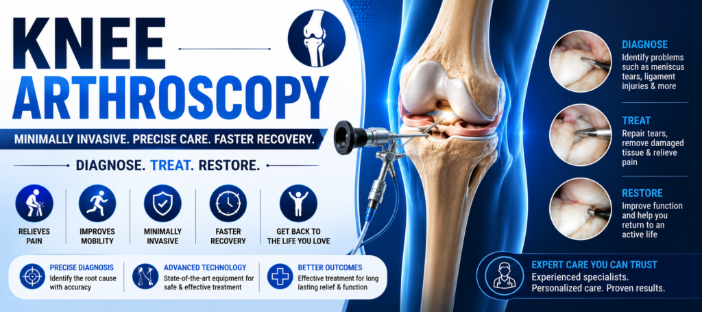
Knee Arthroscopy
Knee Arthroscopy is a minimally invasive surgical procedure that uses a tiny camera called an arthroscope to examine, diagnose, and treat problems inside the knee joint. Small incisions are made around the knee to insert the camera and specialized surgical instruments.
What is Knee Arthroscopy?
During the procedure, a miniature camera is inserted into the knee joint, allowing the surgeon to view the internal structures on a high-definition monitor. Arthroscopy enables accurate diagnosis and treatment while causing minimal damage to surrounding tissues.
Because of its minimally invasive nature, knee arthroscopy often results in less pain, smaller scars, and faster recovery compared to traditional open surgery.
Minimally Invasive
Small incisions reduce tissue damage and promote quicker healing.
High-Definition Visualization
Advanced camera technology provides a clear view inside the knee.
Accurate Diagnosis
Helps identify cartilage, ligament, and meniscal injuries precisely.
Faster Recovery
Most patients return to daily activities sooner than with open surgery.
Types of Anesthesia Used
- Local Anesthesia: Numbs only the knee area while the patient remains awake.
- Spinal Anesthesia: Blocks sensation below the waist while the patient stays awake.
- General Anesthesia: The patient remains completely asleep during surgery.
- Femoral Nerve Block: Regional anesthesia that reduces pain and anesthesia requirements.
How Knee Arthroscopy is Performed
The surgeon makes two or three tiny incisions around the knee. Sterile saline solution is introduced into the joint to improve visibility. An arthroscope is inserted through one incision while specialized instruments are inserted through others to repair or remove damaged tissue.
After the procedure, the instruments are removed, the incisions are closed with stitches, and a sterile dressing is applied.
Torn Meniscus Repair
Treatment of cartilage tears that cause pain and knee locking.
ACL & PCL Reconstruction
Repair of damaged cruciate ligaments that affect knee stability.
Cartilage Repair
Correction of damaged cartilage and joint surface defects.
Baker’s Cyst Removal
Treatment of fluid-filled swelling behind the knee.
Why Knee Arthroscopy is Performed
- Torn Meniscus (Cartilage Injury)
- ACL or PCL Ligament Tears
- Inflamed Synovium (Joint Lining)
- Patellar (Kneecap) Misalignment
- Baker’s Cyst
- Cartilage Damage
- Certain Knee Fractures
- Loose Bodies Inside the Joint
Recovery & Outcomes
Recovery after simple procedures such as meniscal repair is often rapid, with many patients recovering within 1–2 weeks. For ligament reconstruction procedures such as ACL or PCL surgery, partial weight-bearing is usually allowed after approximately 3 weeks, while full weight-bearing is generally achieved within 6 weeks.
Most patients experience excellent outcomes with significant pain relief, improved mobility, and restoration of knee function.
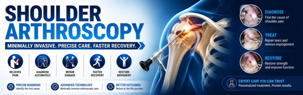
Shoulder Arthroscopy
Shoulder Arthroscopy is a minimally invasive surgical procedure that uses a small camera called an arthroscope to examine, diagnose, and treat problems inside the shoulder joint. The arthroscope is inserted through tiny incisions, allowing surgeons to perform complex repairs with minimal tissue damage and faster recovery.
Understanding the Shoulder & Rotator Cuff
The rotator cuff consists of muscles and tendons that stabilize the shoulder joint and allow smooth movement of the arm in different directions.
Overuse, sports injuries, aging, or trauma can cause tears in the rotator cuff tendons, leading to pain, weakness, and restricted movement.
Minimally Invasive Surgery
Small incisions reduce tissue damage, pain, and scarring.
High-Definition Visualization
A miniature camera provides a clear view inside the shoulder joint.
Accurate Repairs
Damaged tendons, ligaments, cartilage, and tissues can be repaired precisely.
Faster Recovery
Patients typically recover faster than with traditional open surgery.
Common Procedures Performed During Shoulder Arthroscopy
- Rotator Cuff Repair
- Shoulder Impingement Surgery
- Labral Tear Repair
- Bankart Lesion Repair
- SLAP Lesion Repair
- Shoulder Instability Correction
- Removal of Damaged Tissue
- Bone Spur Removal
How Shoulder Arthroscopy is Performed
The procedure is usually performed under general anesthesia or regional anesthesia. The surgeon inserts the arthroscope through a small incision and examines the shoulder joint on a video monitor.
Additional small incisions allow specialized instruments to repair torn tendons, cartilage, ligaments, or remove damaged tissue. After completion, the incisions are closed with stitches and covered with dressings.
Rotator Cuff Repair
Torn tendons are repaired and secured to the bone using suture anchors.
Impingement Syndrome Surgery
Inflamed tissue and bone spurs are removed to relieve pain.
Bankart Lesion Repair
Repair of the damaged cartilage rim in shoulder instability cases.
SLAP Lesion Repair
Treatment of injuries affecting the upper labrum and ligament complex.
Possible Risks & Complications
- Bleeding
- Blood Clots
- Infection
- Shoulder Stiffness
- Persistent Pain
- Failure of Tendon Healing
- Shoulder Weakness
- Nerve or Blood Vessel Injury
After the Procedure
Patients are provided with detailed discharge and rehabilitation instructions. Pain medications may be prescribed to improve comfort during recovery.
Most patients will need to wear a shoulder sling for at least one week. More extensive repairs may require a longer period of immobilization.
Recovery & Outlook
Recovery usually takes between 1 to 3 months, depending on the type of repair performed. Physical therapy plays a critical role in restoring shoulder strength, mobility, and function.
Arthroscopic shoulder surgery has excellent outcomes for rotator cuff tears, shoulder instability, labral injuries, and impingement syndrome. Most patients experience significant pain relief and improved shoulder function.
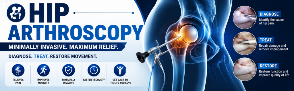
Hip Arthroscopy
Hip Arthroscopy is a minimally invasive surgical procedure used to diagnose and treat a variety of conditions affecting the hip joint. Although it is less commonly performed than knee or shoulder arthroscopy, advancements in surgical techniques have made it an effective treatment option for many hip disorders.
What is Hip Arthroscopy?
Hip arthroscopy involves inserting a small camera called an arthroscope through tiny incisions around the hip joint. The camera projects images onto a monitor, allowing the surgeon to accurately diagnose and treat problems inside and around the hip.
Initially used mainly for diagnosing unexplained hip pain, hip arthroscopy is now widely used to manage both intra-articular and extra-articular hip conditions with minimal tissue damage and faster recovery.
Minimally Invasive
Small incisions lead to less pain, reduced scarring, and quicker recovery.
Accurate Diagnosis
Provides a direct view of the hip joint for precise evaluation.
Advanced Treatment
Enables treatment of complex hip disorders without major surgery.
Faster Rehabilitation
Most patients experience quicker recovery compared to open procedures.
Common Conditions Treated with Hip Arthroscopy
- Femoroacetabular Impingement (FAI)
- Labral Tears
- Loose Body / Foreign Body Removal
- Hip Joint Infection (Hip Washout)
- Hip Biopsy Procedures
- Cartilage (Chondral) Lesions
- Osteochondritis Dissecans
- Ligamentum Teres Injuries
- Ligamentum Teres Reconstruction
Advanced Hip Conditions Managed Arthroscopically
Modern hip arthroscopy techniques allow surgeons to address several soft tissue and tendon-related disorders affecting the hip region.
Iliopsoas Tendinopathy
Treatment of painful snapping hip syndrome involving the psoas tendon.
Trochanteric Pain Syndrome
Management of chronic pain affecting the outer side of the hip.
Snapping Iliotibial Band
Relief from painful snapping sensations around the hip joint.
Sciatic Nerve Compression
Treatment of conditions such as Piriformis Syndrome causing nerve irritation.
Additional Indications
- Ischiofemoral Impingement
- Selected Cases of Osteoarthritis
- Direct Assessment of Hip Replacement Implants
- Persistent Unexplained Hip Pain
- Joint Preservation Procedures
- Sports-Related Hip Injuries
Benefits of Hip Arthroscopy
Hip arthroscopy has become an important advancement in orthopedic surgery, offering patients effective treatment options with shorter recovery times, improved joint function, and excellent long-term outcomes.